
Morpho-anatomy of Adventitious Roots of Vanilla (Vanilla planifolia Jacks. ex Andrews) During Attachment to Support Post
Keywords:
vanilla, adventitious roots, attachmentAbstract
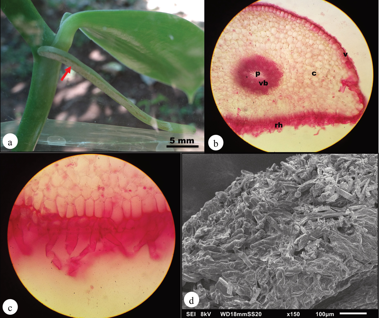
The attachment strategy of vanilla adventitious roots was characterized into three stages according to the size and the extent of adhesion, namely: (1) initial contact; (2) weak to strong attachment and; (3) strong and permanent attachment. The root tip was identified to send signals to produce profuse unicellular elongated root hairs in the velamen on the ventral side of the root facing the substrate which interacted with the surface of the substrate that brought the adhesion. Strong and irreversible attachment was observed between roots and textured substrate.References
Bowling, A.J. and K.C. Vaughn. 2009. Gelatinous fibers are widespread in coiling tendrils and twining vines. American Journal of Botany 96(4): 719–727.
Franceschi, V.R. and P.A. Nakata. 2005. Calcium oxalate in plants: Formation and function. Annual Reviewof Plant Biology 56: 41–71.
Kausch, A.P. and H.T. Horner. 1983. Development of syncytial raphide crystal idioblasts in the cortex of adventitious roots of Vanillaplanifolia L. (Orchidaceae). Scanning Electron Microscopy II: 893–903.
Lenaghan, S.C. and M. Zhang. 2012. Real-time observation of the secretion of a nanocomposite adhesive from English ivy (Hedera helix). PlantScience 183: 206–211. development and morphology of the attachment system of Parthenocissus tricuspidata. InternationalJournal of Plant Sciences 172(9): 1120–1129.
Stern, W.L. and W.S. Judd. 1999. Comparative vegetative anatomy and systematics of Vanilla (Orchidaceae). Botanical Journal of the LinneanSociety 131: 353–382.
Vaughn K.C. and A.J. Bowling. 2011. Biology andphysiology of vines. In: Janick J. (ed.) Horticultural Reviews, 38. Wiley-Blackwell, pp. 1–21.
Downloads
Published
How to Cite
Issue
Section
License
Copyright (c) 2020 National Science Museum, Thailand

This work is licensed under a Creative Commons Attribution-NonCommercial-NoDerivatives 4.0 International License.


