
Histology and Ultrastructure of the Vitelline Gland of the Pancreatic Fluke 𝙀𝙪𝙧𝙮𝙩𝙧𝙚𝙢𝙖 𝙥𝙖𝙣𝙘𝙧𝙚𝙖𝙩𝙞𝙘𝙪𝙢
Keywords:
vitelline gland, trematode, eggshell formation, 𝘌𝘶𝘳𝘺𝘵𝘳𝘦𝘮𝘢 𝘱𝘢𝘯𝘤𝘳𝘦𝘢𝘵𝘪𝘤𝘶𝘮Abstract
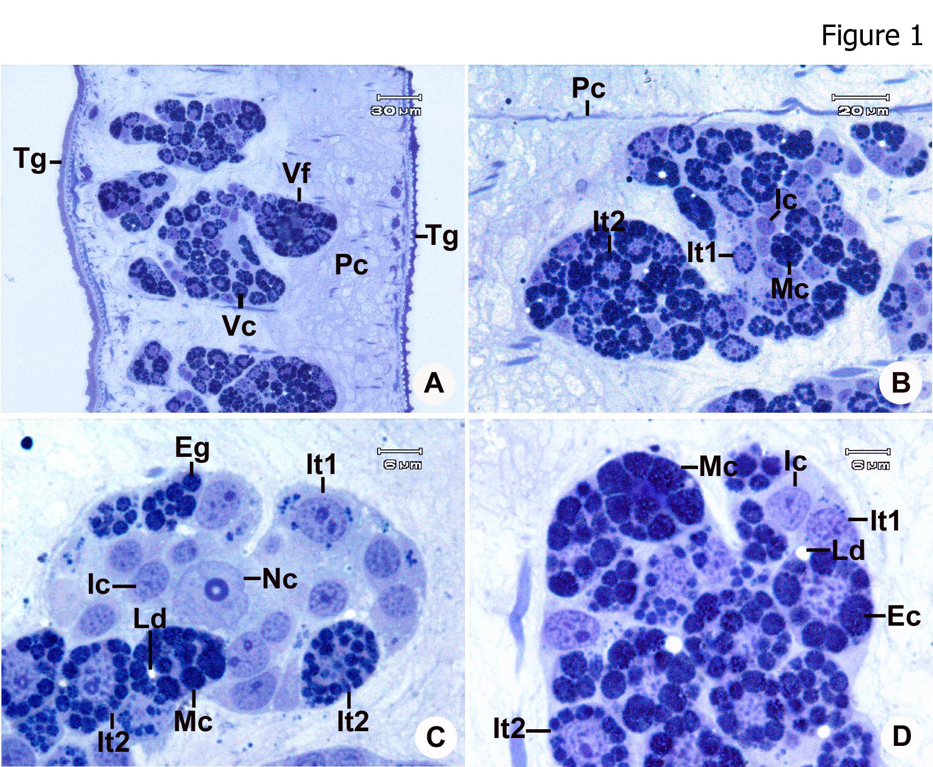
Background and Objectives: The vitelline gland is an essential reproductive structure in trematodes, with vitelline cells contributing to eggshell formation and providing nutrients and other components required for embryonic development. During vitellogenesis, developing vitelline cells undergo a series of morphological and ultrastructural changes associated with the synthesis and accumulation of specific cytoplasmic inclusions, particularly eggshell globules and lipid droplets. The composition and organization of these inclusions vary among digenean species and may reflect differences in reproductive strategies and patterns of egg development. In trematodes that produce embryonated eggs, vitelline cells may exhibit specialized structural characteristics associated with embryonic development prior to egg release from the definitive host. However, detailed information on the ultrastructural differentiation of vitelline cells in trematodes that produce embryonated eggs remains limited. In particular, the structural relationship between developing vitelline cells and their associated nurse cells has not been extensively characterized. To address this gap, this study investigated the histological organization and ultrastructure of the vitelline gland of Eurytrema pancreaticum, an embryonated egg-producing digenean trematode, with particular emphasis on the sequential differentiation of vitelline cells and the structural characteristics of nurse cells during vitellogenesis.
Methodology: Vitelline glands from five adult E. pancreaticum specimens were processed for resin embedding and examined using light microscopy (LM) and transmission electron microscopy (TEM). Semithin sections were examined by LM to characterize the organization and distribution of vitelline follicles. Ultrathin sections were examined by TEM to characterize the ultrastructural features of vitelline cells at different developmental stages and their associated nurse cells. Vitelline cells were classified into developmental stages based on their cytoplasmic organization and the presence, size, and distribution of characteristic cytoplasmic inclusions. Ultrastructural assessment focused oneggshell globule clusters, lipid droplets, glycogen particles, glycan vesicles, cellular junctions, and the association between vitelline cells and nurse cells.
Main Results: The vitelline gland consisted of numerous follicles arranged in two lateral groups on either side of the parasite. Each follicle contained developing vitelline cells surrounded by flattened nurse cells. Based on their ultrastructural characteristics, vitelline cells were classified into four developmental stages: immature, intermediate type I, intermediate type II, and mature cells. Immature vitelline cells exhibited relatively undifferentiated cytoplasm with early development of characteristic inclusions. During intermediate differentiation, eggshell globules progressively appeared, increased in number and size, and became organized into distinct clusters. Lipid droplets also accumulated progressively throughout development. Intermediate type II cells showed more prominent eggshell globule clusters and increased lipid accumulation, indicating further cellular differentiation. Mature vitelline cells contained large, well-developed eggshell globule clusters and abundant lipid droplets, representing the most advanced stage of vitelline cell differentiation. In contrast, glycogen particles and glycan vesicles were not observed at any developmental stage examined. Nurse cells exhibited extensive apical invaginations and close associations with adjacent developing vitelline cells. Gap junctions were observed between nurse cells and developing vitelline cells, suggesting intercellular communication and functional interactions during vitellogenesis. The coordinated development of eggshell globules and lipid inclusions, together with the close association between nurse cells and developing vitelline cells, indicates that vitelline cell differentiation is an organized process involving interactions between developing and supporting cells.
Conclusions: This study provides detailed histological and ultrastructural information on vitelline cell differentiation in E. pancreaticum. Vitellogenesis was characterized by the progressive formation and enlargement of eggshell globule clusters and the accumulation of lipid droplets, whereas glycogen particles and glycan vesicles were absent throughout the developmental sequence. The extensive apical invaginations and gap junctions observed between nurse cells and developing vitelline cells further indicate close structural and functional interactions during vitellogenesis. The distinctive combination of eggshell globule development, lipid accumulation, and nurse cell association may be related to the reproductive strategy of this embryonated egg-producing trematode. These findings contribute to the understanding of reproductive biology in E. pancreaticum and provide useful ultrastructural information for comparative studies of vitellogenesis and eggshell formation among digenean trematodes.
References
Awad, A.H., & Probert, A.J. (1990). Ultrastructure and histochemistry of vitelline cells of Schistosoma mansoni. Journal of Helminthology, 64, 229–239.
Erasmus, D.A. (1975). An ultrastructural study of the vitelline cell of Schistosoma mansoni. Parasitology, 70, 303–318.
Irwin, S.W.B., & Threadgold, L.T. (1970). Electron microscope studies of Fasciola hepatica. II. The vitelline cells. Parasitology, 61, 321–330.
Irwin, S.W.B., & Threadgold, L.T. (1972). The ultrastructure of the vitelline cells of the liver fluke Fasciola hepatica. Parasitology, 64, 49–59.
Meepool, A., Wanichanon, C., Viyanant, V., & Sobhon, P. (2006). Development and roles of vitelline cells in eggshell formation in Fasciola gigantica. Invertebrate Reproduction and Development, 49, 9–17.
Pang, D. H., Shen, H., & Ni, Y. H. (1991). Ultrastructural study on the vitelline cells of Pagumogonimus skrjabini. Zhongguo Ji Sheng Chong Xue Yu Ji Sheng Chong Bing Za Zhi, 9(4), 281–283.
Rao, K.H. (1958). Studies on the vitellaria of trematodes. Proceedings of the Indian Academy of Sciences Section B, 48, 1–10.
Ruangsittichai, J., Viyanant, V., Vichasri-Grams, S., Sobhon, P., Tesana, S., Upatham, E.S., Hofmann, A., Korge, G., & Grams, R. (2006). Opisthorchis viverrini: Identification of a glycine–tyrosine rich eggshell protein and its potential as a diagnostic tool for human opisthorchiasis. International Journal for Parasitology, 36, 1329–1339.
Schmidt, G.D. (1998). Foundations of Parasitology (6th ed.). Boston: McGraw-Hill.
Seed, J.R., & Bennett, J.L. (1978). Biochemical aspects of schistosome eggshell tanning. Experimental Parasitology, 44, 33–42.
Świderski Z, Kacem H, Mackiewicz JS,& Miquel J. (2019). Functional ultrastructure and cytochemistry of vitellogenesis and mature vitellocytes of the digenean Cainocreadium labracis. Parasitology Research 118,493–504.
Threadgold, L.T. (1982). Fasciola hepatica: A scanning and transmission electron microscope study. London: Academic Press.
Ualiyeva RM, Zhangazin SB, & Altayeva IB. (2022). Structural Organization of Vitelline Cells of Trematode Azygia lucii. OnLine Journal of Biological Sciences, 22,10–17.
Wells, K.E., & Cordingley, J.S. (1991). Structure and cross-linking of schistosome eggshell proteins. Parasitology, 103, 351–360.
Downloads
Published
How to Cite
Issue
Section
License
Copyright (c) 2026 Faculty of Science, Burapha University

This work is licensed under a Creative Commons Attribution-NonCommercial-NoDerivatives 4.0 International License.
Burapha Science Journal is licensed under a Creative Commons Attribution-NonCommercial-NoDerivatives 4.0 International (CC BY-NC-ND 4.0) licence, unless otherwise stated. Please read our Policies page for more information