
Comparative Evaluation of EDTA-Based Surface Decalcification Efficiency in Paraffin-Embedded Femoral Bone Blocks of Mlac:NZW Rabbits
Keywords:
rabbits, femur , decalcification, decalcifying agents, EDTAAbstract
Background and Objectives: Histopathological examination of bone tissue requires an effective decalcification process because calcium, the principal mineral component of bone, impedes sectioning by microtomy at the standard thickness of approximately 4 µm. The presence of residual mineralized tissue can adversely affect specimen preparation. However, sectioning artifacts were still observed, affecting tissue integrity and slide quality. These artifacts may compromise microscopic evaluation and reduce the diagnostic quality of histological sections. In routine histopathology laboratories, bone specimens are commonly decalcified using ethylenediaminetetraacetic acid (EDTA) prior to tissue processing and paraffin embedding. EDTA is a calcium ions chelating agent that removes mineralized components while preserving tissue morphology and staining quality more effectively than acidic decalcifying agents. In addition, EDTA is particularly suitable for bone marrow studies because it causes minimal alteration of lymphoid surface antigens and provides superior preservation of nucleic acid integrity, including messenger RNA (mRNA). Although conventional EDTA decalcification is generally performed for 7–14 days and is considered effective for the removal of calcium deposits, some paraffin-embedded bone specimens remain difficult to section. This observation suggests that residual mineral components may persist within the tissue despite prolonged decalcification, thereby affecting microtomy performance and section quality. Such challenges may lead to increased workload, repeated sectioning attempts, and delayed histopathological evaluation. Despite these advantages, the optimal duration of EDTA treatment required to improve the sectioning quality of previously processed bone paraffin blocks has not been clearly established. Surface decalcification of paraffin blocks has been proposed as a practical and time-efficient alternative technique for improving microtomy performance in incompletely decalcified bone specimens. This approach involves exposing the block surface to a decalcifying solution immediately before sectioning, thereby targeting residual mineral deposits without repeating the entire tissue-processing procedure. Therefore, the present study aimed to determine the optimal duration of EDTA surface decalcification for improving the histological quality of femoral bone sections obtained from Mlac:NZW rabbits. The femur was selected as the experimental model because it is a dense long bone with substantial mineral content, making it suitable for evaluating the effectiveness of surface decalcification. The findings of this study may contribute to the development of standardized protocols for surface decalcification and improve the efficiency and quality of bone tissue preparation for routine histopathological examination.
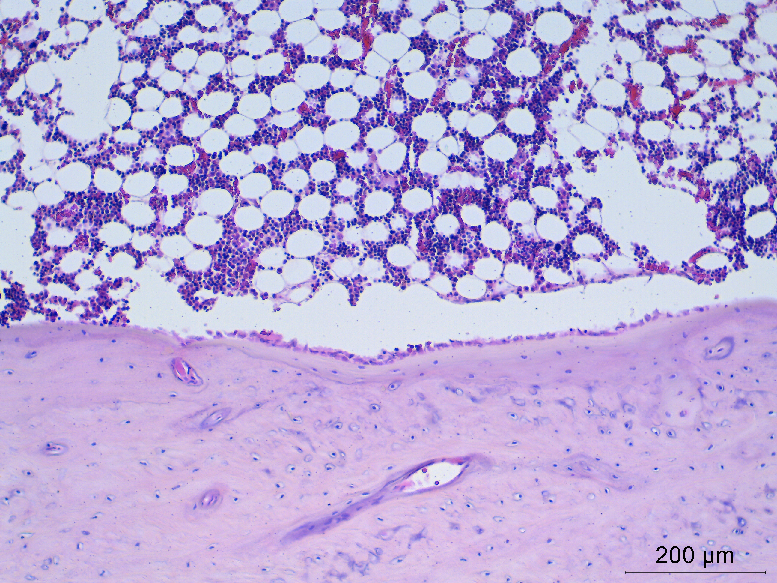
Methodology: Twenty-four femoral bone specimens obtained from New Zealand White rabbits (Mlac:NZW) were fixed in 10% neutral buffered formalin for 48 h and decalcified in EDTA solution for 24 h at room temperature (25°C) prior to routine histological tissue processing. The paraffin blocks were randomly assigned to four groups (n = 6 per group) according to the duration of EDTA surface decalcification: 0 min (control), 15 min, 30 min, and 45 min. Following treatment, tissue sections were cut at a thickness of 4 µm, stained with hematoxylin and eosin (H&E), and evaluated for sectioning ease and histological slide quality.
Main Results: Surface decalcification of paraffin blocks with EDTA improved both sectioning ease and permanent slide quality compared with the untreated control group. Sectioning ease increased with longer EDTA immersion times (15, 30, and 45 min). The 30-min EDTA treatment produced the highest overall H-score, demonstrating optimal preservation of tissue architecture and H&E staining quality, and differed significantly from the control and 45-min treatment groups (P ≤ 0.05). Although the 45-min treatment further facilitated sectioning, it showed a tendency toward reduced slide quality compared with the 30-min treatment. The 15-min treatment improved sectioning performance and slide quality compared with the control group but yielded lower overall H-scores than the 30-min and 45-min treatment groups.
Conclusions: Surface decalcification of paraffin blocks using EDTA for 30 min was identified as the optimal duration for preparing femoral bone tissue from Mlac:NZW rabbits. This method may serve as a practical approach for improving bone tissue preparation and enhancing the efficiency of routine histopathological procedures.
References
Alers, J. C., Krijtenburg, P.-J., Vissers, K. J., & van Dekken, H. (1999). Effect of bone decalcification procedures on DNA in situ hybridization and comparative genomic hybridization: EDTA is highly preferable to a routinely used acid decalcifier. Journal of Histochemistry & Cytochemistry, 47(5), 703-709. https://doi.org/10.1177/002215549904700512
Bogoevski, K., Woloszyk, A., Blackwood, K., Woodruff, M. A., & Glatt, V. (2019). Tissue morphology and antigenicity in mouse and rat tibia: Comparing 12 different decalcification conditions. Journal of Histochemistry & Cytochemistry, 67(8), 545–561. https://doi.org/10.1369/0022155419850099
Bumalee, D., & Meesakul, O. (2017). The comparative study of the effectiveness of decalcifying agents on human jaws. Mahidol Dental Journal, 37(3), 327-338. (in Thai)
Choube, A., Astekar, M., Choube, A., Sapra, G., Agarwal, A., & Rana, A. (2018). Comparison of decalcifying agents and techniques for human dental tissues. Biotechnic & Histochemistry, 93(2), 99–108.
Chow, D. H., Zheng, L., Tian, L., Ho, K.-S., Qin, L., & Guo, X. (2019). Application of ultrasound accelerates the decalcification process of bone matrix without affecting histological and immunohistochemical analysis. Journal of Orthopaedic Translation, 17, 112–120.
Kang, Q. K., LaBreck, J. C., Gruber, H. E., & An, Y. H. (2003). Histological techniques for decalcified bone and cartilage. Handbook of histology methods for bone and cartilage. https://doi.org/10.1007/978-1-59259-417-7_13
Kaweblum, M., Aguilar, M. C., Blancas, E., Kaweblum, J., Lehman, W. B., Grant, A. D., & Strongwater, A. M. (1994). Histological and radiographic determination of the age of physeal closure of the distal femur, proximal tibia, and proximal fibula of the New Zealand white rabbit. Journal of Orthopaedic Research, 12(5), 747–749. https://doi.org/10.1002/jor.1100120519
Li, Z., Wenhart, C., Reimann, A., Cho, Y.-L., Adler, K., & Muench, G. (2024). Hypertonic saline-and detergent-accelerated EDTA-based decalcification better preserves mRNA of bones. Scientific Reports, 14(1), 1-18. https://doi.org/10.1038/s41598-024-61459-8
Sanjai, K., Kumarswamy, J., Patil, A., Papaiah, L., Jayaram, S., & Krishnan, L. (2012). Evaluation and comparison of decalcification agents on human teeth. Journal of Oral and Maxillofacial Pathology, 16(2), 222-227. https://doi.org/10.4103/0973-029X.99070
Skinner, R. A. (2003). Decalcification of bone tissue. Handbook of histology methods for bone and cartilage. https://doi.org/10.1007/978-1-59259-417-7_10
Sterchi, D. L. (2019). Bone. Bancroft's Theory and Practice of Histological Techniques, Edition 8.
Suvarna, S. K., Layton, C., & Bancroft, J. D. (Eds.). (2013). Paraffin section cutting. Bancroft's theory and practice of histological techniques, Edition 7.
Walsh, L., Freemont, A. J., & Hoyland, J. A. (1993). The effect of tissue decalcification on mRNA retention within bone for in situ hybridization studies. International Journal of Experimental Pathology, 74(3), 237–241.
Downloads
Published
How to Cite
Issue
Section
License
Copyright (c) 2026 Faculty of Science, Burapha University

This work is licensed under a Creative Commons Attribution-NonCommercial-NoDerivatives 4.0 International License.
Burapha Science Journal is licensed under a Creative Commons Attribution-NonCommercial-NoDerivatives 4.0 International (CC BY-NC-ND 4.0) licence, unless otherwise stated. Please read our Policies page for more information