
Leaf Epidermal Anatomy of Medicinal Plants in Lamtakhong Research Station, Nakhon Ratchasima Province
Keywords:
leaf epidermal , qualitative anatomy , medicinal plants , Lamtakhong Research StationAbstract
Background and Objectives : Nowadays, medicinal plants are being utilized in a wide range of applications, leading to the compilation of traditional knowledge and local wisdom from various sources within Nakhon Ratchasima Province. However, accurate species identification prior to use is crucial, as some plants may exhibit similar external morphological features while possessing distinct anatomical structures that influence phytochemical accumulation and medicinal properties. Leaf epidermal anatomy of medicinal plants serves as an important diagnostic tool because these characters are genetically inherited, can be examined throughout all seasons, and allow species identification even when plant materials are incomplete, highly fragmented, or ground into herbal powders, conditions that increase the likelihood of adulteration with parts from different plant species. The aim of this study was therefore to establish a reference database of leaf epidermal anatomical characteristics for selected medicinal plant species found in the herbal garden of the Lamtakhong Research Station, Nong Sarai Subdistrict, Pak Chong District, Nakhon Ratchasima Province. This research station is a significant learning resource in Thailand, housing a diverse collection of medicinal plants used in both pharmaceutical applications and pest control. Nevertheless, anatomical information on the leaf epidermis of many species cultivated in this garden remains lacking. The findings from this study help fill this knowledge gap and provide foundational data that can be applied to future work in plant taxonomy and pharmacobotany.
Methodology : Medicinal plants were gathered from the herbal garden at Lamtakhong Research Station, Nakhon Ratchasima Province, and fixed in 70% ethyl alcohol for preservation. Anatomical characteristics of the leaf epidermis were examined for 86 medicinal plant species, representing 75 genera and 42 families. The characters investigated included cuticular ornamentation, stomatal types and position, stomatal density, epidermal cell shape and density, trichome types and distribution patterns, and the presence of crystals and other inclusions. The specimens were prepared by the clearing technique using a 5 percent sodium hydroxide (NaOH) solution. The mature leaf blades (margin, blade, and midrib) were cut into sections of approximately 5×5 mm. The samples were immersed in a 5% Clorox solution for 15-30 minutes (until the sample is colorless). Afterward, rinse with clean water 4–5 times. Dehydration was performed using a graded ethanol series (30%, 50%, and 70%) for 15 minutes each, respectively. The samples were stained with 1% Safranin-O in 70% in ethyl alcohol solution for 3–12 hours, then washed off the excess stain with 95% ethanol 2–3 times. Dehydrated with a series of increasing 95% ethanol, 100% ethanol, then a 1:1 mixture of ethanol and xylene, and finally in pure xylene, for 30 minutes at each step. The slides were mounted using DePeX. The abaxial and adaxial surfaces of leaf epidermis were described under the light microscope (LM). Photographs were taken using a Leica ICC50W digital camera connected to the light microscope. The voucher specimens were deposited in the Herbarium of Nakhon Ratchasima Rajabhat University.
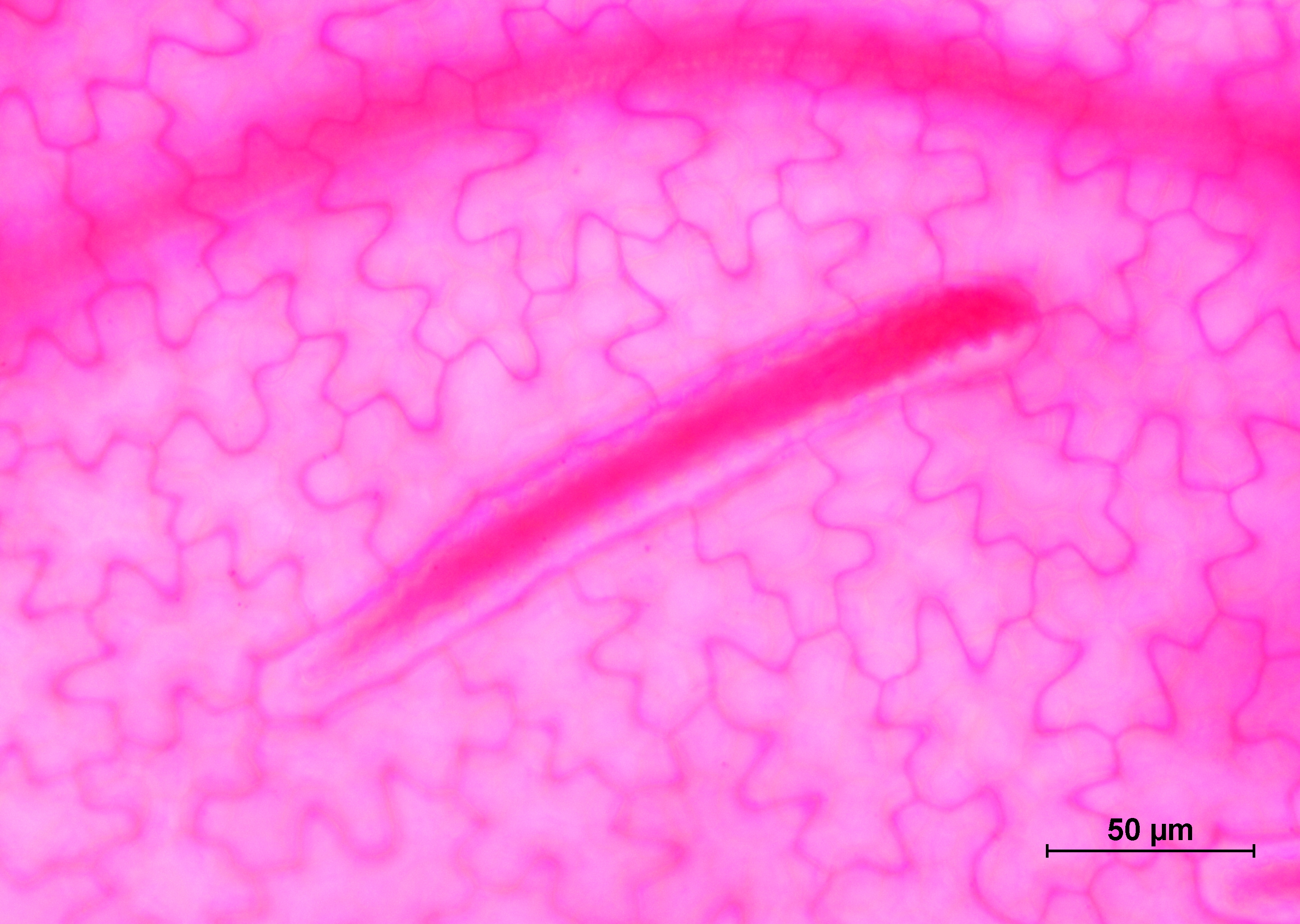
Main Results : The generalized qualitative anatomical characteristics of the eighty six medicinal plants studied were as follows: 1) the cuticular ornamentation is smooth or striated 2) the shapes of epidermal cells are polygonal, jigsaw-like and irregular, 3) the stomata are present in both abaxial and adaxial epidermis (amphistomatic leaf) or present in only abaxial epidermis (hypostomatic leaf), the stomatal types are anomocytic, paracytic, actinocytic, cyclocytic, tetracytic, anisocytic and diacytic, 4) the trichome are present in both sides, only abaxial or absent, the trichome types are unicellular trichome, bicellular trichome, multicellular trichome and glandular trichome, and 5) the types of inclusions are prismatic crystal, druse crystal, raphide crystal, sand crystal cystolith crystal and inclusion not present. These characteristics can be used to identify some closely related species. The quantitative data of leaf epidermal anatomical characteristics in adaxial surface: Trevesia palmata had the highest average epidermal cell density (236.55±11.0 cell/mm2) while Dracaena angustifolia had the lowest average epidermal cell density 33.44±4.19 cell/mm2). Murraya paniculata had the highest average stomatal density (60.33±7.12 stomata/mm2) while Platostoma palustre had the lowest average stomatal density (1.11±0.31 stomata/mm2). The quantitative of leaf epidermal anatomical data in abaxial surface: Buddleja paniculata had the highest average epidermal cell density (185.00±6.63 cell/mm2) while Strobilanthes cusia had the lowest average epidermal cell density (7.44 ±11.28 cell/mm2). Murraya paniculata had the highest average stomatal density (63.44±9.87 stomata/mm2) while Platostoma palustre had the lowest average stomatal density (2.33±1.33 stomata/mm2).
Conclusions : The leaf epidermal anatomical study of medicinal plant species at Lamtakhong Research Station revealed several qualitative anatomy features that can be used to authenticate certain species present in the area. These include variation in cuticular ornamentation patterns, shape of epidermal cells, stomatal cells position, type of stomata, type of trichomes, and crystal type and deposit. Quantitative anatomical data are variable and cannot be used to identify all medicinal plants; however, they can be used to distinguish between certain species that share similar qualitative characteristics. Such information provides a fundamental reference framework that can support future applications in the pharmacognostic and pharmaceutical evaluation of medicinal plants.
References
Department of Agriculture. (2014). Medicinal plants, thai wisdom. Bangkok: Printing House of the Agricultural Cooperative Federation of Thailand Limited. (in Thai)
Gardner, S., Sidisunthorn, P., & Anusarnsunthorn, V. (2007). A field guide to forest trees of northern Thailand. Bangkok, Thailand: Kobfai Publishing Project. (in Thai)
Kesonbua, W., & Kodkaew, Y. (2022). Leaf epidermal anatomy for the identification of some medicinal plants in Ubon Ratchathani University. Journal of Science and Technology, Ubon Ratchathani University, 24(1), 17-33. (in Thai)
Khonkayan, S., Lamyai, S., Tanawet, K., & Machana, K. (2021). Leaf epidermal anatomy of some species of the family Fabaceae in the northeastern of Thailand. Journal of Roi Et Rajabhat University: Science and Technology, 2(1), 26-34. (in Thai)
Lersten, M.R., & Curtis, J.D. (2001). Idioblast and other unusual internal folia secretory structures in Scrophulariaceae. Plant Systematics and Evolution, 227, 63-73.
Metcalfe, & Chalk. (1979). Anatomy of the dicotyledons: Vol. I, systematic anatomy of leaf and stem, with a briefhistory of the subjects. 2nd edition. Oxford: Clarendon Press.
Nakhon Ratchasima Provincial Cultural Office. (2006). Korat city history, a record of oral traditions from local wisdom, Nakhon Ratchasima: Sombun Printing. (in Thai)
Phonsena, P. (2007). Medicinal plants in Khao Hin Son herbal forest garden, complete edition. Chachoengsao, Thailand: Forest Botany Division, Khao Hin Sorn Royal Development Study Center. (in Thai)
Phonsena, P. (2013). Medicinal plants in botanical garden and arboretum in Thailand. Bangkok, Thailand: Research Office for Forest Conservation and Plant Diversity, Department of National Parks, Wildlife and Plant Conservation. (in Thai)
Phonsena, P. (2015). Medicinal plants in botanical garden and arboretum in Thailand 2. Bangkok, Thailand: Research Office for Forest Conservation and Plant Diversity, Department of National Parks, Wildlife and Plant Conservation. (in Thai)
Songnok, T., & Channete, N. (2018). Local wisdom in treatment of traditional chemists in Nakhon Ratchasima. NRRU Community Research Journal, 12(3), 124-135. (in Thai)
Suthanon, T., Srinual, A.,& Kesonbua, W. (2006) Leaf anatomy of some medicinal plants in mangrove forest of Eastern Thailand. Thai Journal of Botany, 8(2), 307-325. (in Thai)
Downloads
Published
How to Cite
Issue
Section
License
Copyright (c) 2026 Faculty of Science, Burapha University

This work is licensed under a Creative Commons Attribution-NonCommercial-NoDerivatives 4.0 International License.
Burapha Science Journal is licensed under a Creative Commons Attribution-NonCommercial-NoDerivatives 4.0 International (CC BY-NC-ND 4.0) licence, unless otherwise stated. Please read our Policies page for more information