Effects of Mercury(II) Chloride and Osil® (OsilR) on Node Surface Sterilization of Hom (Strobilanthes cusia (Nees) Kuntze) for Initiating In Vitro Axenic Plantlets
Keywords:
Strobilanthes cusia , surface sterilization , in vitro culture , mercury(II) chloride , silver nanoparticlesAbstract
Background and Objectives : Hom (Family : Acanthaceae, Scientific name : Strobilanthes cusia (Nees) Kuntze) is a plant producing an indigo compound that is essential for dyeing in the Morhom textile industry. Ensuring an adequate supply of Hom for dye extraction is crucial, therefore plant tissue culture techniques are employed to propagate this plant effectively. However, the mass propagation of Hom through tissue culture requires a substantial number of axenic plantlets. As a consequence, effective surface sterilization of explants is imperative for initiating in vitro contamination-free plantlets. A proper surface disinfection procedure will verify the rapid implementation of Hom tissue culture by establishing a large quantity of starting material for plant multiplication. Previous studies have examined the surface sterilization of Hom explants using sodium hypochlorite (NaOCl). While NaOCl proved effective in eliminating contamination, it induced tissue damage and cell rupture, resulting in the leakage of indigo pigment. Consequently, when these explants were cultured under aseptic tissue culture conditions, they failed to develop and ultimately died. Thus, exploring alternative chemical agents for surface sterilization of Hom explants warrants further investigation, as these agents may enhance sterilization efficiency while reducing tissue damage and explant mortality. Several studies on plant tissue culture in the Acanthaceae family have indicated that mercury(II) chloride (HgCl2) provides a high rate of both disinfection and survival of explants. Moreover, recent research has explored the application of silver nanoparticles (AgNPs) in surface sterilization processes. These nanoparticles have demonstrated effective antimicrobial properties while maintaining a high survival rate of the explants. Therefore, the objective of this study is to develop and improve the surface sterilization process of Hom explants by evaluating the effectiveness of surface sterilization using HgCl2 and Osil® (OsilR), which is an agricultural chemical and its active ingredient for eliminating microorganisms is AgNPs at 0.01% w/w.
Methodology : Healthy Hom plants, exhibiting no visible signs of disease or insect damage and possessing stem heights of approximately 40–50 cm, were selected for excision of 3–4 cm long nodal segments to be used as experimental plant material. The explants were subjected to surface sterilization using either 0.1% and 0.2% (w/v) HgCl2 solutions or 10% and 15% (v/v) OsilR solutions, with soaking durations of 10 or 15 minutes, resulting in a total of eight treatment combinations. Each treatment consisted of 10 explants and was replicated three times. The nodes were subsequently cultured on Murashige & Skoog (MS) medium to evaluate the effectiveness of the surface sterilization process. After four weeks of culture, the contamination-free rate of the explants was recorded. The sterile explants were then transferred to fresh MS medium and cultured for an additional four weeks to assess their survival rate, resulting in a total culture period of eight weeks.
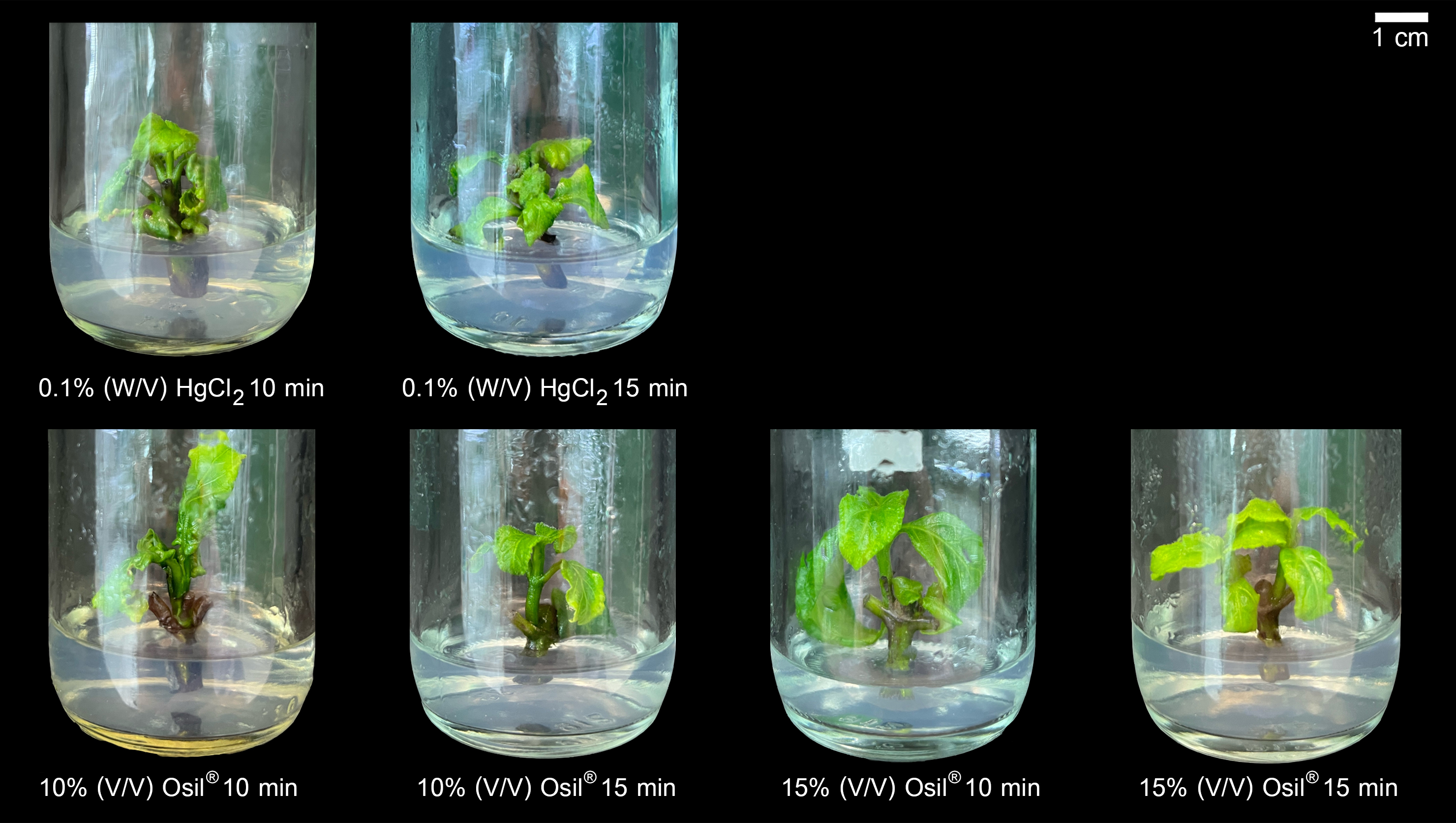
Main Results : Surface sterilization of Hom nodal segments using HgCl2 solution resulted in an average contamination-free rate of 78.33%. In comparison, disinfection with OsilR solution provided a higher average contamination-free rate of 90.00%. Increasing both the chemical concentration and the exposure time was found to significantly enhance sterilization efficiency. Notably, treatment with 0.2% (w/v) HgCl2 solution for 15 minutes completely eliminated microbial contamination. Similarly, disinfection using 15% (v/v) OsilR solution for 10 or 15 minutes achieved a 93.33% contamination-free rate in nodal explants. The surface sterilization methods employed in this study did not cause observable damage to the explants, as evidenced by the absence of indigo pigment release. However, different surface sterilization treatments resulted in varying survival rates among contamination-free explants. Nodal segments sterilized with HgCl2 showed an overall survival rate of 28.37%, while those disinfected with OsilR exhibited a significantly higher survival rate of 95.35%. Although increasing the concentration of HgCl2 and AgNPs, along with prolonged exposure time, improved decontamination efficiency, these conditions significantly reduced explant viability. Specifically, treatment with 0.2% (w/v) HgCl2 solution for 10 or 15 minutes led to complete mortality of axenic explants. Surface sterilization of Hom nodes using 0.1% (w/v) HgCl2 solution for 10 minutes resulted in a survival rate of 64.44%, which was the highest among all explants disinfected with HgCl2 solution. In contrast, nodal segments sterilized with 10% (v/v) OsilR solution for 15 minutes or 15% (v/v) OsilR solution for 10 minutes achieved a 100% survival rate among contamination-free explants. The lowest survival rate within the OsilR-treated groups (85.56%) was observed in explants disinfected with 15% (v/v) OsilR solution for 15 minutes.
Conclusions : Surface sterilization of Hom nodal segments using 15% (v/v) OsilR solution for 10 minutes was identified as the most effective method in this study. This treatment provided a high contamination-free rate of 93.33%, with 100% survival of all sterilized explants. The findings from this research can be applied to enhance the efficiency of surface sterilization protocols for Hom explants, enabling the achievement of a greater number of contamination-free and viable explants. OsilR may be considered a promising alternative chemical for the surface sterilization of Hom explants prior to introduction into tissue culture systems, thereby improving the overall potential for Hom propagation through plant tissue culture techniques.
References
Abdi, G., Salehi, H., & Khosh-Khui, M. (2008). Nano silver: a novel nanomaterial for removal of bacterial contaminants in valerian (Valeriana officinalis L.) tissue culture. Acta Physiologiae Plantarum, 30, 709-714.
Ahlawat, J., Sehrawat, A.R., Choudhary, R., & Yadav, S.K. (2022). Biologically synthesized silver nanoparticles eclipse fungal and bacterial contamination in micropropagation of Capparis decidua (FORSK.) Edgew: A substitute to toxic substances. Indian Journal of Experimental Biology, 58(5), 336-343.
Andújar, I., González, N., García-Ramos, J.C., Bogdanchikova, N., Pestryakov, A., Escalona, M., & Concepción, O. (2020). Argovit™ silver nanoparticles reduce contamination levels and improve morphological growth in the in vitro culture of Psidium friedrichsthalianum (O. Berg) Nied. SN Applied Sciences, 2, 2110.doi.org/10.1007/s42452-020-03948-9.
Arab, M.M., Yadollahi, A., Hosseini-Mazinani, M., & Bagheri, S. (2014). Effects of antimicrobial activity of silver nanoparticles on in vitro establishment of G X N15 (hybrid of almond X peach) rootstock. Journal of Genetic Engineering & Biotechnology, 12(2), 103-110.
Asnawi, A., Hartinie, M., & Jualang, A.G. (2017). Effect of cytokinins on shoot induction from nodal explant of Pecah Beling (Strobilanthes crispus). In 26th Malaysian Society of Plant Physiology Conference. (pp. 8-12). Malaysia : Sarawak.
Böhme, M., Diener, M., Mestres, P., & Rummel, W. (1992). Direct and indirect actions of HgCl2 and methyl mercury chloride on permeability and chloride secretion across the rat colonic mucosa. Toxicology & Applied Pharmacology, 114(2), 285-294.
Boruah, J. (2020). Effect of 0.1% HgCl2 on surface sterilization of Som (Persea bombycina King) explant during tissue culture — A major host plant of Muga silkworm. International Journal of Current Microbiology & Applied Sciences, 9, 954-958.
Bucio, L., García, C., Souza, V., Hernández, E., González, C., Betancourt, M., & Gutiérrez-Ruiz, M.C. (1999). Uptake, cellular distribution and DNA damage produced by mercuric chloride in a human fetal hepatic cell line. Mutation Research/Fundamental and Molecular Mechanisms of Mutagenesis, 423(1–2), 65-72.
Chaiai, P., Sangsoy, V., Putivoranat, M., Charoenkid, S., Suriyapromchai, P., & Konchom, R. (2021). Increase of the production potential of Strobilanthes cusia (Nees) for local textile dyeing in the upper north of Thailand. Thai Agricultural Research Journal, 39(1), 96-109. (in Thai)
Choengpanya, K., Susawaengsup, C., Sornsakdanuphap, J., Dangtungee, R., & Siangsuepchart, A. (2024). Antifungal property of silver nano-chito oligomer hybrid solution against canbendazim-resistant fungus. Fusarium solani. In The 4th National and The 2nd International MJU-Phrae Conference. (pp. 60-66). Thailand: Phare.
Cuong, D.M., Mai, N.T.N., Tung, H.T., Khai, H.D., Luan, V.Q., Phong, T.H., Van The Vinh, B., Phuong, H.T.N., Van Binh, N., & Tan Nhut, D. (2023). Positive effect of silver nanoparticles in micropropagation of Limonium sinuatum (L.) Mill.‘White’. Plant Cell, Tissue & Organ Culture, 155(2), 417-432.
Fukuzaki, S. (2006). Mechanisms of actions of sodium hypochlorite in cleaning and disinfection processes. Biocontrol Science, 11(4), 147-157.
Gammoudi, N., Nagaz, K., & Ferchichi, A. (2022). Establishment of optimized in vitro disinfection protocol of Pistacia vera L. explants mediated a computational approach: multilayer perceptron–multi–objective genetic algorithm. BMC Plant Biology, 22, 324. doi.org/10.1186/s12870-022-03674-x.
Gwaltney-Brant, S.M. (2023). Chapter 10 - Metals. In W.M. Haschek, C.G. Rousseaux, M.A. Wallig, & B. Bolon. (Eds.), Haschek and Rousseaux' s Handbook of Toxicologic Pathology Volume 3 : Environmental Toxicologic Pathology and Selected Toxicant Classes. (pp. 679-725). Cambridge: Academic Press.
Hashim, S.N., Ghazali, S.Z., Sidik, N.J., Chia-Chay, T., & Saleh, A. (2021). Surface sterilization method for reducing contamination of Clinacanthus nutans nodal explants intended for in-vitro culture. E3S Web of Conferences, 306, 01004. doi:10.1051/e3sconf/202130601004
Ho, Y.L., Kao, K.C., Tsai, H.Y., Chueh, F.Y., & Chang, Y.S. (2003). Evaluation of antinociceptive, anti-inflammatory and antipyretic effects of Strobilanthes cusia leaf extract in male mice and rats. American Journal of Chinese Medicine, 31(1), 61-69.
Joseph, S. (2014). In Vitro Propagation, Phytochemical And Biological Evaluation of Strobilanthes Cuspidata (Benth) T. Anderson (Ph.D. Thesis). Tamilnadu : Department of Botany, School of Life Science, Bharathiar University.
Khaldoun, K., Khizar, S., Saidi-Besbes, S., Zine, N., Errachid, A., & Elaissari, A. (2024). Synthesis of silver nanoparticles as an antimicrobial mediator. Journal of Umm Al-Qura University for Applied Sciences. doi : 10.1007/s43994-024-00159-5
Khanam, B.,& Chandra, R. (2017). Optimization of surface sterilization process of selected dye-yielding plants for isolation of bacterial endophytes. In Applications of Biotechnology for Sustainable Development. (pp. 45-50). Singapore: Singapore.
Krupa-Malkiewicz, M., Oszmianski, J., Lachowicz, S., Szczepanek, M., Jaskiewicz, B., Pachnowska, K., & Ochmian, I. (2019). Effect of nanosilver (nAg) on disinfection, growth, and chemical composition of young barley leaves under in vitro conditions. Journal of Integrative Agriculture, 18(8), 1871-1881.
Leva, A., & Rinaldi, M.R. (2012). Recent Advances in Plant In Vitro Culture. Rijeka : InTechOpen.
Mahajan, S., Kadam, J., Dhawal, P., Barve, S., & Kakodkar, S. (2022). Application of silver nanoparticles in in-vitro plant growth and metabolite production: revisiting its scope and feasibility. Plant Cell, Tissue & Organ Culture, 150(1), 15-39.
Mahna, N., Vahed, S.Z., & Khani, S. (2013). Plant in vitro culture goes nano: nanosilver-mediated decontamination of ex vitro explants. Journal of Nanomedicine & Nanotechnology, 4, 2. doi : 10.4172/2157-7439.1000161
Mangkita, W., & Lattirasuvan, T. (2013). Ecological characteristics and production of Baphicacanthus cusia (Ness) Brem. at Phrae Province. Thai Agriculture Research Journal, 31(1), 26-40. (in Thai)
Mangkita, W., Wongmuang, S., & Panngom, K. (2011). Micropropagation of Hom (Strobilanthes cusia (Nees) Kuntze). Thai Journal of Botany, 3(2), 187-197. (in Thai).
Murashige, T., & Skoog, F. (1962). A revised medium for rapid growth and bio assays with tobacco tissue cultures. Physiologia Plantarum, 15(3), 473-497.
Sarmast, M., Salehi, H., & Khosh-Khui, M. (2011). Nano silver treatment is effective in reducing bacterial contaminations of Araucaria excelsa R. Br. var. Glauca explants. Acta Biologica Hungarica, 62(4), 477-484.
Sekerli, M. S. (2024). Season, thermotherapy and surface sterilization play important roles in microbial contamination of hazelnut in vitro cultures. Plant Cell, Tissue & Organ Culture, 157(3), 70. doi : 10.1007/s11240-024-02799-1
Shameer, M.C., Saeeda, V.P., Madhusoodanan, P.V., & Sailas Benjamin, S.B. (2008). In vitro propagation of Strobilanthes hamiltoniana. Journal of Tropical Medicinal Plants, 9(1), 77-81.
Srikun, N. (2017). In vitro propagation of the aromatic herb Strobilanthes tonkinensis Lindau. Agriculture & Natural Resources, 51(1), 15-19.
Susawaengsup, C., Choengpanya, K., Sornsakdanuphap, J., Tabtimmai, L., Chaiharn, M., & Bhuyar, P. (2023). Phytochemical and pharmacological properties of a traditional herb, Strobilanthes cusia (Nees) Kuntze. Molecular Biotechnology. doi : 10.1007/s12033-023-00897-7
Tanaka, T., Ikeda, T., Kaku, M., Zhu, X.H., Okawa, M., Yokomizo, K., Uyeda, M., & Nohara, T. (2004). A new lignan glycoside and phenylethanoid glycosides from Strobilanthes cusia Bremek. Chemical & Pharmaceutical Bulletin, 52(10), 1242-1245.
Vatcharakajon, P., Sornsaket, A., Choengpanya, K., Susawaengsup, C., Sornsakdanuphap, J., Boonplod, N., Bhuyar, P., & Dangtungee, R. (2023). Silver nanochito oligomer hybrid solution for the treatment of Citrus greening disease (CGD) and biostimulants in Citrus. Horticulture, 9(6), 725. doi: 10.3390/horticulturae 9060725.
Wang, L., Hu, C., & Shao, L. (2017). The antimicrobial activity of nanoparticles: present situation and prospects for the future. International Journal of Nanomedicine, 12, 1227-1249.
Zhang, Y., Chen, Y.Y., Huang, L., Chai, Z.G., Shen, L.J., & Xiao, Y. H. (2017). The antifungal effects and mechanical properties of silver bromide/cationic polymer nano-composite-modified Poly-methyl methacrylate-based dental resin. Scientific reports, 7(1), 1547. doi : 10.1038/s41598-017-01686-4
Downloads
Published
How to Cite
Issue
Section
License
Copyright (c) 2025 Faculty of Science, Burapha University

This work is licensed under a Creative Commons Attribution-NonCommercial-NoDerivatives 4.0 International License.
Burapha Science Journal is licensed under a Creative Commons Attribution-NonCommercial-NoDerivatives 4.0 International (CC BY-NC-ND 4.0) licence, unless otherwise stated. Please read our Policies page for more information